Ganglion Cell
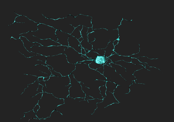
Retinal ganglion cells (RGCs) are the cells that are responsible for transferring information from the eye to the brain. These cells come with distinct functional signature, size, and morphology, and their dendritic arbors have been shown to be confined to specific sublayers of the inner plexiform layer (IPL) of the retina. However, despite this heterogeneity in structure, connectivity and function, to this point, many studies on the development of the visual system and its pathways have regarded all RGC's as being part of a single group. Studies are now emerging that show that different subclasses of RGC's are maximally responsive to particular visual features, and that they arborize selectively within particular layers of the IPL. The number of RGC subtypes and the extent to which they differ from one another remains to be seen, but based upon studies on dendritic morphology, it is estimated that, in mammals, there are approximately 20 subtypes of RCG's.
Contents
Introduction
A retinal ganglion cell is a type of neuron located near the inner surface (the ganglion cell layer) of the retina of the eye. It receives visual information from photoreceptors via two intermediate neuron types: bipolar cells and amacrine cells. Retinal ganglion cells collectively transmit image-forming and non-image forming visual information from the retina to several regions in the thalamus, hypothalamus, and mesencephalon, or midbrain.
Retinal ganglion cells vary significantly in terms of their size, connections, and responses to visual stimulation but they all share the defining property of having a long axon that extends into the brain. These axons form the optic nerve, optic chiasm, and optic tract. A small percentage of retinal ganglion cells contribute little or nothing to vision, but are themselves photosensitive; their axons form the retinohypothalamic tract and contribute to circadian rhythms and pupillary light reflex, the resizing of the pupil.
Physiology
Ganglion cells are the final output neurons of the vertebrate retina. The ganglion cell collects the electrical messages concerning the visual signal from the two layers of nerve cells preceding it in the retinal wiring scheme. A great deal of preprocessing has been accomplished by the neurons of the vertical pathways (photoreceptor to bipolar to ganglion cell chain), and by the lateral pathways (photoreceptor to horizontal cell to bipolar to amacrine to ganglion cell chain) before presentation to the ganglion cell and so it represents the ultimate signaller to the brain of retinal information. Ganglion cells are larger on average than most preceding retinal interneurons and have large diameter axons capable of passing the electrical signal, in the form of transient spike trains, to the retinal recipient areas of the brain many millimeters or centimeters distant from the retina. The optic nerve collects all the axons of the ganglion cells and this bundle of more than a million fibers (in humans at least) then passes information to the next relay station in the brain for sorting and integrating into further information processing channels.
Anatomy
Location
The ganglion cells lie innermost in the retina closest to the lens and front of the eye.
Connections
Ganglion cells extract features of the visual world, encode them in frequency modulated spike trains, and send them coursing along optic nerve axons to various visual brain centers. The first step in this process is the binding of amacrine and bipolar cell neurotransmitters onto specialized receptor proteins embedded in ganglion cell dendritic membrane. These receptor molecules are concentrated at sites of synapses with amacrine and bipolar cells. Once the neurotransmitter is bound, ion selective channels, a kind of micropore in the subsynaptic membrane, open. Charged ionic species rush through these channels, driven by electrochemical gradients. As a consequence current flows into the cell altering membrane potential. These synaptic currents, through a complex interplay of dendritic structure and other potential sensitive ion channels (Fohlmeister and Miller, 1997), alter ganglion cell firing rates, and convey visual signals to the brain. The axons of ganglion cells that are located in the retina are not myelinated, while axons that are located outside the retina are myelinated.
Molecules
Retinal ganglion cells respond to all common excitatory or inhibitory retinal neurotransmitters. When neurotransmitters are applied to the solution bathing ganglion cells, membrane currents are induced. AMPA, kainate or NMDA evoke excitatory currents in both ON and OFF type cat beta cells (Cohen et al, 1994). NMDA currents are of the typical ‘conditional’ sort, dominant only if cells are depolarized first by other excitatory neurotransmitters, or in the absence of extracellular magnesium. Extracellular acetylcholine also excites retinal ganglion cells (Masland and Ames, 1976; Lipton et al, 1987; Cohen et al, 1994).
The bath applied, inhibitory retinal neurotransmitters GABA and glycine potently evoke inhibitory currents in ganglion cells. GABA receptors in cat beta cells are of the ‘A’ type. These responses can be largely blocked by the GABA antagonist bicuculline. Strychnine effectively blocks glycine-evoked currents (Cohen et al, 1994).
Ganglion cell membranes appear to be broadly tuned to receive signals from many neurotransmitter systems. Rather than being neurotransmitter selective, they limit input of information from amacrine and bipolar cells by selective synaptic connections. Thus it is important to study the properties of responses evoked by natural photic stimulation to determine which of the available transmitter systems in the retinal inner plexiform layer are actually utilized. In extracellular impulse recordings, metabotropic glutamate receptors were shown important for the ON center circuit in cat alpha and beta ganglion cells, whereas both these receptors and glycine receptors were found important for OFF alpha and beta cells (Müller et al,1988).
History

Cajal in his monumental work on Golgi staining of the vertebrate retina was able to classify many different varieties of ganglion cell based on form (dendritic morphology), extent (cell body and dendritic tree size), and number of sublayers in which they arborize (stratification levels in the inner plexiform layer). He considered the retina to be remarkably uniform across all vertebrates differing only in respect to rod and cone specializations for the visual sense of the animal. Looking at Cajals (1892) drawings of dog ganglion cells as compared frog ganglion cell for example the former seem simpler in form than the latter cells but we actually now know that there is a common evolutionary path taken by different ganglion cell types so that different morphological and functional classes are similar throughout the species. For example the large ganglion cells, with open radiate branching patterns, process fast, transient impulse trains and in all vertebrate retinas are concerned with motion detection and alerting the animal to threatening, moving visual imagery. While small bushy ganglion cell types are concerned with processing small stationary, fine detail in tonically activated messages in all species.
In the forties, Polyak (1941) produced a phenomenal description of the Golgi-impregnated neurons of the primate retina and therein he gave us a good classification of ganglion cell types. So by the sixties we had a fairly extensive description and classification of the ganglion cells in mammalian and monkey retinas but all the data was based on vertical sections of stained ganglion cells (Cajal, 1892; Polyak, 1941; Brown and Major, 1966; Leicester and Stone, 1967; Boycott and Dowling, 1969; Shkolnik-Yarros, 1971). The advent of a technique to perform Golgi staining on wholemount retinas allowed a reinterpretation of many of the earlier classifications, because now we could see the entire dendritic tree of a ganglion cell.
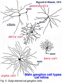
A particularly successful morphological classification scheme was proposed by Boycott and Wassle (1974). Three main classes of physiological response type were correlated with three morphological classes of ganglion cell in cat retina. Thus the alpha, beta, gamma and delta morphological ganglion cell types were considered to be the equivalents of the Y, X and W types of the physiology (Boycott and Wassle, 1974; Enroth-Cugell and Robson, 1966; Cleland and Levick, 1974, Levick and Thibos, 1983, for a review). By comparison with Cajals drawings, it was clear that alpha and beta types were already described (see Cajal drawing of dog and ox retinal ganglion cells) and a further ten or more of Cajals (1892) types were lumped under the umbrella of gamma and delta groups.
By 1978, we knew that alpha and beta cells of Boycott and Wassle (1974) could be subdivided into separate subtypes depending on whether they branch in sublamina a or sublamina b of the inner plexiform layer (Famiglietti and Kolb, 1976) (see chapter on inner plexiform layer). Sublamina a contained dendrites of cells with physiologically defined OFF-center receptive fields and sublamina b dendrites of cell with ON-center receptive fields (Nelson et al., 1978). A new classification of Golgi stained ganglion cells (Kolb et al., 1981) could now relate both alpha and beta cells to the functionally important sublamina scheme and further, described many new types of cell including and going beyond the gamma and delta cells. The 20 cell types over and above the alpha, beta and gamma cells were named from G4-G23 in order of cell body size and dendritic form (i.e. from smallest types, G4 to largest types at G23) (Kolb et al., 1981).
References
- ↑ Kolb, Helga, Nelson, Ralph, Fernandez, Eduardo, Jones, Bryan, The Organization of the Retina and Visual System, Simple Anatomy of the Retina. url=http://webvision.med.utah.edu/book/part-i-foundations/simple-anatomy-of-the-retina/


