Latest revision as of 18:32, 14 June 2016
Information about message (contribute ) This message has no documentation.
If you know where or how this message is used, you can help other translators by adding documentation to this message.
Message definition (E2198 )
[[Image:e2198_sbem.png|thumb|c|left|280px|Image of the retina from serial block-face scanning electron microscopy (SBFSEM). Scale bar = 100 microns.]]
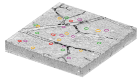
[[Image:e2198_sbem.png|thumb|c|left|280px|Image of the retina from serial block-face scanning electron microscopy (SBFSEM). Scale bar = 100 microns.]]
Immediately after 2P imaging, the retina was fixed, stained, and embedded in a hard plastic resin. An unconventional stain was used to mark the boundaries between neurons while leaving intracellular organelles unstained. SBFSEM was used to image a volume of size 350×300×60 µm{{math|<sup>3</sup>}} (left) with voxel resolution 16.5×16.5×23 nm{{math|<sup>3</sup>}}. Since the same blood vessels were visible in both the 2P and SBFSEM images, the researchers were able to find the same DSGCs in both images. These are marked by colored circles on the SBFSEM image just as in the 2P image above.
Translation [[Image:e2198_sbem.png|thumb|c|left|280px|Image of the retina from serial block-face scanning electron microscopy (SBFSEM). Scale bar = 100 microns.]] Image of the retina from serial block-face scanning electron microscopy (SBFSEM). Scale bar = 100 microns.
Immediately after 2P imaging, the retina was fixed, stained, and embedded in a hard plastic resin. An unconventional stain was used to mark the boundaries between neurons while leaving intracellular organelles unstained. SBFSEM was used to image a volume of size 350×300×60 µm3 3
