Difference between revisions of "The Eye and Retina/ko"
(Created page with "신경절 세포들은 망막에서 빛을 처리하는 과정의 맨 마지막 단계에 해당합니다. 어떤 것이 무슨 색인지를 처리하기 위해 원추세포...") |
(Created page with "====수평세포 피드백(Horizontal Cell Feedback)==== 수평세포가 망막의 작은 부분을 대표하는 세포이지만, 광수용체 세포와 다수의 시냅스를...") |
||
| Line 93: | Line 93: | ||
신경절 세포들은 망막에서 빛을 처리하는 과정의 맨 마지막 단계에 해당합니다. 어떤 것이 무슨 색인지를 처리하기 위해 원추세포로부터의 신호들이 비교되는 것이 바로 신경절 세포에 의해 이루어집니다. 신경절 세포들은 망막에서 두뇌로 신호를 전달합니다.<ref name=\"Masland2001\" /> | 신경절 세포들은 망막에서 빛을 처리하는 과정의 맨 마지막 단계에 해당합니다. 어떤 것이 무슨 색인지를 처리하기 위해 원추세포로부터의 신호들이 비교되는 것이 바로 신경절 세포에 의해 이루어집니다. 신경절 세포들은 망막에서 두뇌로 신호를 전달합니다.<ref name=\"Masland2001\" /> | ||
| − | ====Horizontal Cell Feedback==== | + | ====수평세포 피드백(Horizontal Cell Feedback)==== |
| − | + | 수평세포가 망막의 작은 부분을 대표하는 세포이지만, 광수용체 세포와 다수의 시냅스를 형성합니다. 수평세포의 기능에 대한 유력한 학설 중 하나에 따르면 밝은 부분과 어두운 부분의 대조를 증가시키기 위한 것이라고 합니다. 수평세포들은 흥분된 원추세포의 신호와 주위를 둘러싼 원추세포들을 억제할 것입니다. 이렇게 되면 신호가 약해지는 반면 흥분되지 않은 상태인 주변의 원추세포로부터의 신호 역시 제거하게 되어, 원추세포 및 그 주변 세포와 연관된 모든 신경절 세포들이 흥분되는 것이 아니라 흥분된 상태의 원추세포와 연관된 신경절 세포들만이 흥분되게 됩니다. | |
The rods receive feedback from specialized horizontal cells, which also give feedback to cones, in such a way that the two feedback systems are kept separate.<ref name="Masland2001" /> | The rods receive feedback from specialized horizontal cells, which also give feedback to cones, in such a way that the two feedback systems are kept separate.<ref name="Masland2001" /> | ||
Revision as of 05:07, 8 January 2016
thumb|right|450px|
UNIQaa0a16fd421dd36d-nowiki-00000001-QINU1UNIQaa0a16fd421dd36d-nowiki-00000002-QINU
눈은 다른 감각들에 비해, 우리를 둘러싼 세상에 대해 비교할 수 없는 수준의 지각을 가능케 해주는 시각의 기관입니다. 눈은 주변 환경으로부터 광자(photon)을 수집하고 광자를 전기적 자극으로 바꿔주며 이렇게 바뀐 자극은 시각적으로 인지되는 영상으로 바뀌게 됩니다. 시각적 처리의 일부는 눈 안에서 행해지지만 대부분은 두뇌의 시각 피질(visual cortex)에서 이뤄지게 됩니다.
Contents
눈의 해부학적 구조(The Anatomy of the Eye)
각막(The Cornea)
각막은 동공과 홍채를 덮고 있는 눈의 투명한 표면입니다. 각막은 단순히 눈을 보호하는 기능뿐만 아니라 빛이 망막으로 가기 위해 지나가는 첫 번째 굴절 표면입니다. 건강한 각막은 혈관이 없으며 따라서 건강한 상태를 유지하기 위해 공기 중으로부터 산소를 받아들입니다. 각막에 산소공급이 오랜 기간 중단되면(가령 콘택트 렌즈를 허용 기간 이상 착용하는 경우), 인체는 각막에 혈관을 자라게 해 이러한 산소 부족을 보충하려 합니다. 이는 부정적인 면역 시스템 반응의 확률을 높이게 됩니다(충혈, 통증, 부종, 결국에는 각막의 영구 손상 등을 유발).
각막은 여섯 겹으로 이루어져 있으며 각각 상피세포, 보우만층, 기질, 두아층, 데스메막, 그리고 각막 내피세포층입니다(The epithelium, bowman's layer, stroma, dua's layer, Descemet membrane and corneal endothelium). [2]
상피세포층은 나머지 각막을 보호하는 역할을 하며 보우만 층 역시 마찬가지 입니다.[3], 기질층은 각막 두께의 90% 가량을 차지하며 투명합니다. 두아 층은 각막의 층들 중 가장 최근에 발견된 층으로 2013년도에 개별적인 층일 가능성이 보고 되었으며,[4] 아직 그 기능은 잘 알려지지 않았습니다. 데스메막과 내피세포 층은 각막과 눈의 나머지 부분들 간의 체액과 영영분 흐름을 조절하는 기능을 합니다.[5][6]
홍채와 동공(The Iris and the Pupil)
파일:300px-Eye_dilate.gif 홍채는 각막과 렌즈 사이에 위치한 눈의 일부분 입니다. 홍채의 중심에는 빛이 눈으로 들어올 수 있게 해주는 조리개인 동공이 있습니다. 홍채의 근육은 밝은 빛에 노출될 때 동공을 축소시키며 어두운 곳에서는 동공을 확대시키는 역할을 합니다. 멜라닌 색소의 정도가 홍채의 색깔을 결정하는 가장 중요한 요소 중 하나입니다. 멜라닌이 상대적으로 적은 경우, 홍채는 푸르거나 초록빛을 띠며 멜라닌이 많은 경우 홍채는 갈색 또는 검은색을 띄게 됩니다.[7]
렌즈(The Lens)
렌즈는 홍채 뒤에 있는 구조물로 빛을 모아 망막에 맺히게 합니다. 성인의 렌즈는 혈관이 없으며 수양액(aqueous humor)로부터 영양분을 받습니다. 그러나 발생단계의 자라나는 렌즈는, 보통 태어나기 전에 퇴행하는, 유리체 동맥(hyaloid artery)으로부터 영양을 공급받습니다. 모양체근이 렌즈의 모양을 바꾸어 빛이 렌즈를 통과해 망막에 맺힐 수 있도록 해주며 이러한 과정을 통해 눈이 다양한 거리에 있는 물체에 초점을 맞힐 수 있게 됩니다. [8]
유리액(The Vitreous Humour)
유리액은 걸쭉한, 젤 같은 액체로 렌즈와 각막 그리고 시신경 원판 사이를 채워 눈의 형태를 유지시켜 주는 역할을 합니다. 이 유리액은 눈 부피의 약 80%를 차지하며 98%는 물로 구성되어 있습니다.[9]
공막(Sclera)
공막은 흔히 눈의 흰자위로 알려져 있습니다. 공막은 흰색의 섬유질 층으로 눈의 앞쪽 부위에서는 투명하게 되어 각막을 형성합니다. 인간의 눈은 홍채가 작아 그 위치가 공막을 배경으로 할 때 어디에 있는지가 명확하다는 점에서 드문 경우입니다. 이러한 특징으로 인해 다른 사람이 어디를 보고 있는지를 구별할 수 있으며 이러한 것이 비언어적 의사소통의 한 형태로 발달하게 되었습니다.
인간 망막의 해부학적 구조(Anatomy of the human retina)
thumb|right|400px|The various types of neurons in the mammalian retina.UNIQaa0a16fd421dd36d-nowiki-0000001C-QINU10UNIQaa0a16fd421dd36d-nowiki-0000001D-QINU 망막은 빛에 민감한 조직의 층이며 눈의 뒤쪽 표면을 덮고 있습니다. 시야에서 들어오는 빛은 눈을 통과하여 망막에 맺혀 영상을 만들어 냅니다. 그에 따라 망막에 존재하는 신경세포들이 이 영상을 탐지하게 되고 그에 따라 일련의 생화학적, 전기적 반응이 시작되며 시신경을 통해 결국에는 두뇌에 있는 시각 피질 부위로 전달 됩니다. 이러한 생화학적, 전기적 신호들은 시각의 근간을 제공해 줍니다.
망막의 세포(Cells of the Retina)
광수용체들(Photoreceptors)
- Cone cell en.png
The anatomy of a cone cell
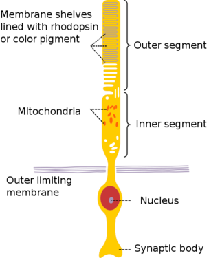
The anatomy of a rod cell[11]

광수용체는 크게 두 가지 종류의 세포, 즉 간상세포와 원추세포로 구성되어 있습니다. 간상세포는 망막의 바깥 쪽 가장자리에 집중되어 있으며 주변 시야에 주로 사용됩니다. 간상세포는 원추세포에 비해 빛에 보다 더 민감하며 (암소시라고도 하는) 야간시력의 거의 전부를 책임지고 있습니다. 원추세포는 망막의 중앙 쪽으로 집중되어 있으며 망막의 중심부위(중심와) 에서 발견되는 유일한 광수용체입니다. 원추세포는 (명소시라고도 하는) 색각을 책임지고 있습니다. 포유류는 보통 둘 또는 세 가지 다른 종류의 원추세포를 가지고 있는 데 이는 자극(예를 들면 색상)의 파장을 명확히 하기 위해서는 적어도 두 가지의 원추세포로부터의 출력 정보가 반드시 비교되어야 하기 때문입니다.
이들 광수용체 세포들은 외절(실제로는 단지 변형된 섬모) 부위에 옵신(opsin)이라는 단백질을 가지고 있으며 이들 옵신이 비타민 A에서 파생된 발색단(chromophore)과 결합해 이들 세포가 흡수된 빛을 전기적 신호로 변환할 수 있게 해줍니다. 빛의 서로 다른 파장은 세포에 발현하는 옵신 단백질의 구조를 변형시켜 감지할 수 있습니다.[12]
수평세포(Horizontal Cell)

수평세포는 두 종류가 존재하는 것을 생각되며 각각의 세포는 구별되는 형태를 가지고 있으며 두 종류 모두 모든 광수용체 세포에 피드백을 제공하는 것으로 간주됩니다. 함께 시냅스를 이루는 세포들의 수와는 상관없이 수평세포들은 망막 세포의 상대적으로 적은 부분만 차지하고 있습니다(내핵층 세포의 5% 미만). 두 종류의 수평세포가 존재하는 정확한 이유는 아직 알려지지 않았습니다; 적-록계에서 색깔을 구분하는 데에 관여할 가능성이 있습니다.
무축삭 세포(Amacrine Cell)

아이와이어에서 재구성된 성화상 무축삭 세포
무축삭 세포는 신경절 세포가 시간적으로 연관된 신호들을 두뇌로 보내도록 해주는 것으로 보입니다: 같은 무축삭 세포로부터 나온 신호가 두 개의 다른 신경절 세포들에 대한 입력 신호로 작용할 때 이들 신경절 세포들은 동시에 신호를 내보내게 되는 경향이 있습니다. 행동이 잘 알려진 무축삭 세포들은 매우 특정한 기능들을 가지고 있는 것으로 나타났습니다.
양극성 세포(Bipolar Cell)

생쥐 망막 조각에서 재구성된 114개의 간상 양극성 신경 세포. 촘촘한 꾸러미(윗 부분) 부분은 수상돌기이며 아래의 듬성듬성한 돌출 부분은 축삭돌기입니다.(사진 제공: 막스플랑크 의학연구소).
양극성 세포들은 광수용체와 신경절 세포를 이어주는 역할을 합니다. 이들의 역할은 광수용체로부터 신경절 세포로, 직접적이던 간접적이던, 신호를 전달하는 것입니다. 양극성 세포는 그 모양에서 이름을 따왔습니다 — 이들 세포는 중심 세포체가 있으며 거기서 두 가지 다른 종류의 신경돌기(축삭 또는 수상돌기)가 뻗어 나옵니다. 이들 세포는 간상 또는 원추 세포와 연결을 형성할 수 있으며(동시에는 불가능), 이들은 또한 수평세포와도 연결을 형성합니다. 활동전위를 이용해 서로 의사소통하는 대다수의 신경세포와는 달리 양극성 세포는 차등전위를 이용해 다른 세포와 “대화”합니다.
신경절 세포(Ganglion Cell)

아이와이어에서 재구성된 신경절 세포
신경절 세포들은 망막의 신호 출력 세포들입니다. 이들 세포의 축삭은 눈을 떠나 시신경을 지나서 두뇌로 이어지며, 처리된 시각적 자극들을 외측 슬상핵(lateral geniculate nucleus)으로 보내고, 자극이 보다 더 해설될 수 있는 부위인 일차 시각 피질로 뻗어 있는 신경세포와 시냅스를 형성합니다.
다 함께 연결해 봅시다.
thumb|left|300px|UNIQaa0a16fd421dd36d-nowiki-00000026-QINU13UNIQaa0a16fd421dd36d-nowiki-00000027-QINU 개별적 세포의 기능을 이해하는 것이 시각을 이해하는 첫 걸음이지만 우리 눈에서 일어난 복잡한 과정들을 센스있게 설명해 주지는 못합니다. 오늘날 과학자들은 시각을 좀더 이해하기 위해서 망막의 커넥톰을 연구하고 있습니다. 그러나 완벽하게 커넥톰을 밝혀내는 것은 현재의 기술로는 매우 어려운 일이며, 대신 과학자들은 부분적 커넥톰을 밝혀내고 있습니다.
신경세포들 간의 연결을 들어다보면 과학자들은 하나의 신경세포에 대한 입력 신호가 연결되어 있는 다른 세포에 어떻게 영향을 미치는가를 이해할 수 있습니다. 과학자들은 다수의 신경세포들 간의 연결들을 마치 하나의 시스템처럼 취급하며 들여다보고 어떻게 하나의 입력 신호가 출력 신호에 영향을 미치는지를 이해하려 애쓰고 있습니다. 과학자들이 한번에 단지 몇몇 신경세포들로 구성된 시스템을 연구하는 반면, 망막이 어떻게 정보를 하나로써 처리하는지에 대한 기본적 이해가 존재합니다.Cite error: Invalid <ref> tag;
refs with no content must have a name
과정의 단순화
가장 기본적인 방식으로 망막의 기능을 설명하기 위해서 광수용체 세포, 양극성 세포, 무축삭 세포, 신경절 세포, 이렇게 네 종류의 망막 신경세포들이 고려될 것입니다. 본 위키 페이지는 보다 많은 망막 신경세포로부터 요인들을 포함시킬 수 있도록 지속적으로 모델을 확장시킬 것입니다.
시각 정보를 처리하는 첫 번째 단계는 광수용체에 의한 광자의 수집입니다. 두 종류의 광수용체, 간상세포와 원추세포는 서로 다른 목적을 갖는 반면 그 기능에서는 유사합니다.
일반적으로 간상세포와 원추세포는 양극성 세포와 시냅스를 형성하게 됩니다. 원추세포의 경우 하나의 원추세포가 하나의 양극성 세포와 시냅스를 형성하는 경우가 많지만 간상세포의 경우에는 다수의 간상세포가 동일한 하나의 양극성 세포와 시냅스를 형성하는 경향이 있습니다. 다수의 간상세포가 하나의 양극성 세포를 공유하기 때문에 영상의 선명함은 신호의 민감도와 맞바꿔집니다.
양극성 세포들은 다음으로 무축삭 세포들과 시냅스를 형성합니다. 무축삭 세포들은 신호 피드백을 제공하며 신호를 신경절 세포에 전달합니다.
신경절 세포들은 망막에서 빛을 처리하는 과정의 맨 마지막 단계에 해당합니다. 어떤 것이 무슨 색인지를 처리하기 위해 원추세포로부터의 신호들이 비교되는 것이 바로 신경절 세포에 의해 이루어집니다. 신경절 세포들은 망막에서 두뇌로 신호를 전달합니다.Cite error: Invalid <ref> tag;
refs with no content must have a name
수평세포 피드백(Horizontal Cell Feedback)
수평세포가 망막의 작은 부분을 대표하는 세포이지만, 광수용체 세포와 다수의 시냅스를 형성합니다. 수평세포의 기능에 대한 유력한 학설 중 하나에 따르면 밝은 부분과 어두운 부분의 대조를 증가시키기 위한 것이라고 합니다. 수평세포들은 흥분된 원추세포의 신호와 주위를 둘러싼 원추세포들을 억제할 것입니다. 이렇게 되면 신호가 약해지는 반면 흥분되지 않은 상태인 주변의 원추세포로부터의 신호 역시 제거하게 되어, 원추세포 및 그 주변 세포와 연관된 모든 신경절 세포들이 흥분되는 것이 아니라 흥분된 상태의 원추세포와 연관된 신경절 세포들만이 흥분되게 됩니다.
The rods receive feedback from specialized horizontal cells, which also give feedback to cones, in such a way that the two feedback systems are kept separate.[14]
Amacrine Cell Feedback
While most cells in the retina are flexible in their function, amacrine cells are highly specialized to do specific tasks.
Scientists have proposed that amacrine cells make ganglion cells fire in correlation with one another, which could increase the amount of information that can be transferred by the optic nerve.[14]
Current Research
According to many neuroscience textbooks, retinal ganglion cells can be categorized into two different types according to a property that is known as their receptive field. Neurons with receptive fields have been found in the auditory (hearing) system, the somatosensory (feeling) system and the visual system, and the receptive field of a particular neuron can generally be defined as a region of space in which the presence of a stimulus will alter the firing of that neuron.
In the visual system, a receptive field of a particular retinal ganglion cell is defined as the region of the photoreceptor cell layer in the retina that alters the firing (signal-sending) of that ganglion cell when it is stimulated with light. According to textbook accounts, retinal ganglion cells either have ON-center, OFF-surround or OFF-center, ON-surround receptive field. An ON-center, OFF-surround ganglion cell will send a signal when the center of its receptive field detects light, but will be inhibited from firing when the area surrounding the center (the surround) of its receptive field detects light; OFF-center, ON-surround cells have the exact opposite response to light stimulation.
Within the last decade, it has become increasingly clear that the notion that only two types of receptive fields exist in photoreceptors is a gross oversimplification. Scientists now know that ganglion cells come in at least 15 or 20 types, each of which has a distinct shape and physiological function, and which correspondingly has connections with different types of cells in the rest of the retina.
At the Max Planck Institute (MPI) for Medical Research in Heidelberg, Germany, a dataset was obtained from a mouse retina in order to investigate this diversity in retinal ganglion cells – by applying two imaging techniques one after the other (two-photon microscopy (2P) and serial block-face scanning electron microscopy (SBFSEM)), scientists have been able to obtain images that show both neural activity and connectivity in retinal ganglion cells. However, the images are very difficult to analyze and interpret, and doing so is a very time-consuming process. Computer scientists at MIT are working on developing software to help with retinal image analysis, but computational analysis is currently much less accurate and reliable than that performed by humans.
The ultimate goal motivating the research on the retina that is being done at places like MPI and MIT is to use 2P and SBFSEM images in order to identify specific cell types within the broad classes of retinal cells that were described earlier, and further to understand connectivity between these cells. Only once the different cell types have been comprehensively catalogued will researchers be able to investigate their specific functions.
This is where YOU come in!
In order to fully understand retinal computation, it is necessary to map all of the connections that converge onto ganglion cells, as this diversity of connections generates the diversity of visual signals that are sent to the brain. The challenge now is to refine the coarse knowledge about retinal connectivity in order to gain a much more in-depth understanding of the specific functions of each and every cell type in the retina. Currently, scientists think that there are at least between fifty and sixty types, so there is much work to be done!
References
- ↑ Eye-diagram no circles border http://commons.wikimedia.org/wiki/File:Eye-diagram_no_circles_border.svg
- ↑ Binder, P. S. et al. (July 1991) High-voltage electron microscopy of normal human cornea. Invest. Ophthalmol. Vis. Sci. 32 (8): 2234-43
- ↑ Dua, Harminder S. et al. (September 2013) Human Corneal Anatomy Redefined: A Novel Pre-Descemet's Layer (Dua's Layer) Ophthalmology 120 (9): 1778-1785 doi: 10.1016/j.ophtha.2013.01.018
- ↑ McKee, Hamish D. et al. (May 2014, published online February 2014) Re: Dua et al.: Human corneal anatomy redefined: a novel pre-Descemet layer (Dua's layer) (Ophthalmology 2013;120:1778–85) Ophthalmology 121 (5): e24-e24 doi: 10.1016/j.ophtha.2013.12.021 \"Abstract: We read the recent claim of the discovery of a new corneal layer by Dua et al with incredulity.1 The existence of pre-Descemet stromal tissue remaining after pneumodissection is well known. Their further investigation of this pre-Descemet stroma confirms that it is stroma, and not a new corneal layer.\"
- ↑ McKee, Hamish D. et al. ANZ Cornea Meeting 2014 Abstracts, page 3 \"Dua’s layer” is just previously described pre-Descemet stroma ... Medical eponyms have traditionally been created by one’s peers to commemorate the importance of a person’s contribution and findings. Dua has taken an interesting step of creating his own eponym, even before his claims have stood the test of further investigation and scrutiny, and despite current trends to avoid medical eponyms (and when they are used, to use the nonpossessive form). If one prefers a medical eponym to describe the pre-Descemet stroma that remains after pneumodissection, then ‘the Feizi stroma’ would be more appropriate.\"
- ↑ Steinert, Roger (Medscape, 01 October 2014) A Controversy in Cornea \"... the purported discovery of a new layer in the cornea ... We all know that today it is quite frowned upon to use names of scientists to describe tissues. It's much more appropriate and helpful to use anatomic terms or physical terms that make sense.\"
- ↑ \"iris.\" Encyclopaedia Britannica. Encyclopaedia Britannica Online Academic Edition. Encyclopædia Britannica Inc., 2014. Web. 16 Jun. 2014. http://www.britannica.com/EBchecked/topic/294031/iris
- ↑ \"lens.\" Encyclopaedia Britannica. Encyclopaedia Britannica Online Academic Edition. Encyclopædia Britannica Inc., 2014. Web. 16 Jun. 2014. http://www.britannica.com/EBchecked/topic/336040/lens
- ↑ \"human eye.\" Encyclopaedia Britannica. Encyclopaedia Britannica Online Academic Edition. Encyclopædia Britannica Inc., 2014. Web. 16 Jun. 2014. http://www.britannica.com/EBchecked/topic/1688997/human-eye/64878/The-transparent-media?anchor=ref531532
- ↑ Masland, R. The fundamental plan of the retina (2001). Nature Neuroscience 4 (9): 877-886
- ↑ Human Physiology and Mechanisms of Disease by Arthur C. Guyton (1992) p.373
- ↑ Plachetzki, D.; Fong, C.; Oakley, T. (2010). \"The evolution of phototransduction from an ancestral cyclic nucleotide gated pathway\". Proceedings. Biological sciences / the Royal Society 277 (1690): 1963–1969. doi:10.1098/rspb.2009.1797. PMC 2880087. PMID 20219739.
- ↑ Kolb, Helga, Nelson, Ralph, Fernandez, Eduardo, Jones, Bryan, The Organization of the Retina and Visual System, Simple Anatomy of the Retina. http://webvision.med.utah.edu/book/part-i-foundations/simple-anatomy-of-the-retina/
- ↑ Cite error: Invalid
<ref>tag; no text was provided for refs namedMasland2001 - ↑ http://en.wikipedia.org/wiki/Receptive_field