Starburst Amacrine Cell
Starburst amacrine cells, also referred to as SAC or SBAC, are a specific subset of amacrine cells present in the retina. Generally speaking, amacrine cells function by affecting the output from bipolar cells. Each of the ~40 subtypes of amacrine cells connects with a particular type of bipolar cell and secretes a particular neurotransmitter. Starburst amacrine cells are noteworthy for being the only cells in the retina to secrete two different kinds of neurotransmitter: the inhibitory neurotransmitter GABA (gamma-aminobutyric acid) as well as the excitatory neurotransmitter acetylcholine (ACh).[2]
Starburst amacrine cells have been shown to preferentially connect to direction-selective ganglion cells (DSGCs), where they provide the main source of inhibition and are essential in the computation of direction selectivity.[3] The asymmetry of the connections between starburst amacrine cells and DSGCs is the prevailing model for how direction selectivity is computed. At least some asymmetry in the connections between these two cell types has been demonstrated,[4] but the exact mechanism of direction selective computation has yet to be determined.
Contents
Physiology
Visual response properties
Cellular biophysics
Anatomy
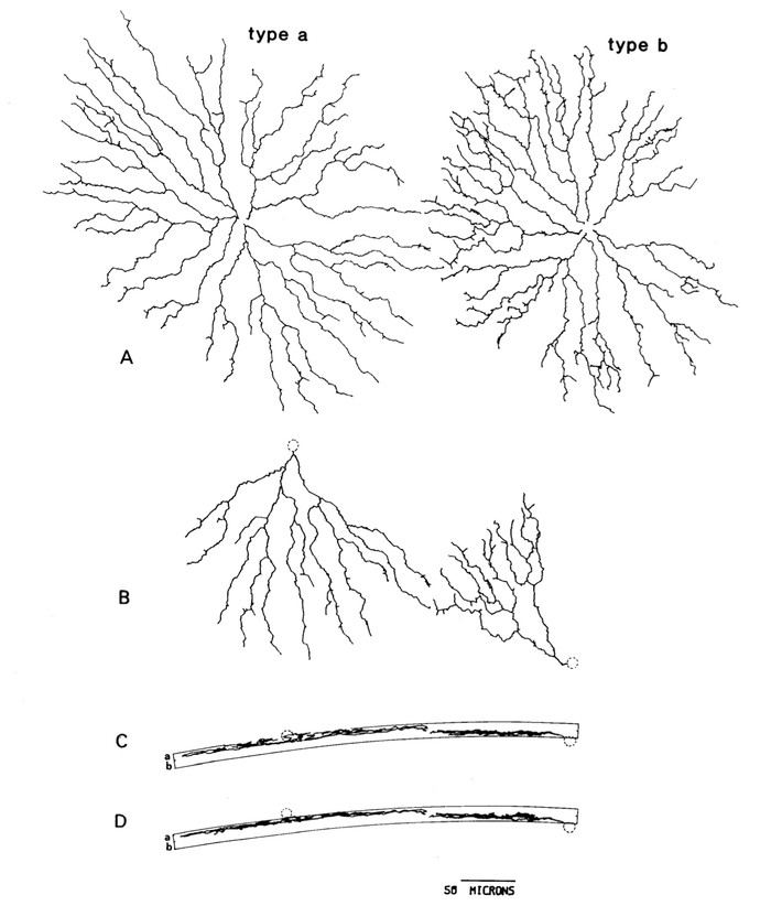
Starburst amacrine cells have a very specific anatomy. Their descriptive name arose from the earliest images of this cell type, in which the characteristic 'starburst' branching of the dendritic arbor could be seen. There are two subytpes of amacrine cells, types a and b. In many categories of cells, classification of subtypes can be inexact and somewhat subjective. For SACs, however, the two subtypes display marked difference in both location, shape, and connections, with these structural differences playing a major role in their function. In fact, it was the distinct difference between subytpes that helped identify starburst amacrine cells as the acetylchole secreting cells in the retina.[5]

Location
The distinct localization of starburst amacrine cell subtypes was fundamental to the identification of SACs as the source of acetylcholine in the retina. By the late 1970s, it was known that acetylcholine (ACh) was synthesized from choline in two different populations of cells in the retina. These ACh synthesizing cells were putatively identified as amacrine cells, since about half of them had cell bodies where one would normally expect to find amacrine cells, flanking the inner plexiform layer (IPL).[6] These were called type a starburst amacrine cells, since they were observed to branch in IPL sublamina a. The other half had their cell bodies in the ganglion cell layer (GCL) but did not appear to be ganglion cells. Since their cell bodies were "displaced" to the ganglion cell layer and they were observed to branch in IPL sublamina b, they are referred to as both type b or displaced starburst amacrine cells.[5] It had previously been shown that starburst amacrine cells also displayed the same localization pattern, and further study confirmed that the starburst amacrine cells were indeed the cholinergic cells in the retina.
There is also a close correlation between location and shape across cell types.
Shape
Starburst amacrine cells are named for the highly distinctive shape of their dendritic arbor.
There are some distinct structural differences between subtypes a and b, several of which can be seen from the figure at the beginning of this section. Generally speaking, the type a cell has a larger dendritic arbor and has a more regular branching pattern.<ref="1985a"></ref> Type a has fewer "branches, spines and boutons in the distal dendritic zone"<ref="1985a"></ref>.
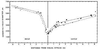
The shape of SACs is also highly correlated to their location in the retina with respect to the visual streak (in rabbits).<ref="1985a"></ref> In humans and some other mammals, the area of highest acuity in the retina is a circular area termed the fovea or area centralis. In contrast, some animals (such as rabbits, the focus of this early SAC study) have a visual streak. In such animals, the area of greatest acuity in their retina is not a single point, but rather an elongated "streak" running across the retina. This allows for better detection of movement in the periphery. In all species studied, the area of highest acuity (whether fovea or visual streak) boasts the highest concentration of cones, the lowest concentration of rods, and much smaller receptive field sizes for all cells. Bipolar cells in this region are smaller, as are ganglion cells. It is unsurprising that starburst amacrine cells also display structural changes as the perpendicular distance from the visual streak increases. Nearest the visual streak, both type a and b SACs have a relatively small cell body size and dendritic field diameter as well as a relatively high frequency of branching and frequency of synaptic boutons.<ref="1985a"></ref> As we move perpendicularly away from the visual streak, cell body size and dendritic field diameter increases, while the frequency of branching and synaptic boutons decreases. (See figure.)
Connections
Molecules
History
In 1976, it was first discovered that there were cells in the rabbit retina that secreted acetylcholine. The most likely candidates were bipolar cells and amacrine cells. Later that same year, it was shown that ganglion cells formed the postsynaptic component of that connection.[7]
Open Questions
References
- ↑ Keeley,P.W.etal.Dendriticspreadandfunctionalcoverageofstarburstamacrine cells. J. Comp. Neurol. 505, 539–546 (2007).
- ↑ O’Malley, D. M., Sandell, J. H. & Masland, R. H. Co-release of acetylcholine and GABA by the starburst amacrine cells. J. Neurosci. 12, 1394–1408 (1992).
- ↑ Yoshida, K. et al. A key role of starburst amacrine cells in originating retinal directional selectivity and optokinetic eye movement. Neuron 30, 771–780 (2001).
- ↑ Briggman, K.L., Helmstaedter, M. & Denk, W. Wiring specificity in the direction-selectivity circuit of the retina. Nature (in the press) (2011).
- ↑ 5.0 5.1 5.2 Famiglietti, E.V. (1985) Starburst amacrine cells: morphological constancy and systematic variation in the anisotropic field of rabbit retinal neurons. J. Neurosci. 5, 562–577
- ↑ Famiglietti, E. V., Jr. (1983) ON and OFF pathways through amacrine cells in mammalian retina: The synaptic connections of “starburst” amacrine cells. Vision Res. 23: 1265-1279.
- ↑ Masland RH, Ames A,, 3rd. Responses to acetylcholine of ganglion cells in an isolated mammalian retina. J Neurophysiol. 1976;39:1220–1235.
