Difference between revisions of "Ganglion Cell/ko"
(Created page with "===연결(Connections)===") |
(Created page with "신경절 세포는 시각적 세상의 특징들을 추출하고 그것을 빈도로 조절되는 spike train 형태로 암호화 하며, 신경 세포 축삭을 따라서...") |
||
| Line 19: | Line 19: | ||
===연결(Connections)=== | ===연결(Connections)=== | ||
| − | + | 신경절 세포는 시각적 세상의 특징들을 추출하고 그것을 빈도로 조절되는 spike train 형태로 암호화 하며, 신경 세포 축삭을 따라서 다시 다양한 두뇌의 시각 센터로 보냅니다. 이러한 과정의 첫 단계는 무축삭 세포와 양극성 세포의 신경전달물질이 신경절 세포 수상돌기막에 박혀있는 특화된 수용체 단백질과 결합하는 것입니다. 이들 수용체 분자들은 무축삭 세포 및 양극성 세포와 시냅스를 형성하는 부위에 집중되어있습니다. 일단 신경전달물질이 결합하면, 아시냅틱막(subsynaptic membrane)에 존재하는 일종의 작은 구멍인 이온 선택적 채널(ion selective channels) 이 열리게 됩니다. 그러면 전기화학적 구배에 의해서 전하를 띄는 이온들이 이들 채널을 통해 쏟아져 들어옵니다. 이러한 결과로, 생체막 전위를 변화시키며 전류가 세포로 흐르게 됩니다. 이러한 시냅스의 전류는, 수지상 구조와 다른 잠재적 민감 이온 채널간의 복잡한 교류작용을 통해서(Fohlmeister and Miller, 1997), 신경절 세포가 발사하는 속도를 변화시키며 시각적 신호를 두뇌로 전달합니다. 망막에 위치한 신경절 세포의 축삭은 막에 덮이지 않은 반면(not myelinated), 망막 외부에 존재하는 신경절 세포의 축삭은 막에 덮여있습니다. | |
==Molecules== | ==Molecules== | ||
Revision as of 19:04, 30 December 2015
망막 신경절 세포(Retinal ganglion cells, RGCs)는 정보를 눈으로부터 두뇌로 전달하는 책임을 지는 세포입니다. 이들 세포들은 독특한 기능적 특징, 크기, 및 형태를 가지고 있으며 이들 세포의 수지상 가지들은 망막 내망상층의 특정한 아층판에만 모여있는 것으로 알려져 있습니다. 그러나 이러한 구조, 연결성 및 기능에서의 다양성에도 불구하고 아직까지는 시각 시스템의 발달과 신호 전달 기전에 관한 많은 연구들이 모든 망막 신경절 세포들을 한 그룹에 속해있는 일부로 간주해왔습니다. 이제 다른 아강(subclass)의 망막 신경절 세포들이 각각 특정한 시각적 기능에 최대로 반응하며 내망상층의 특정한 층에서만 선택적으로 가지를 뻗는 다는 사실을 보여주는 연구들이 나오고 있습니다. 망막 신경절 세포 아강의 수와 서로 얼마나 다른지는 밝혀져야 하겠지만 수지상 돌기의 형태에 대한 연구에 미루어 볼 때 포유류의 경우 약 20개의 망막 신경절 세포 아강이 있는 것으로 추산됩니다.
Contents
도입(Introduction)
망막 신경절 세포는 눈의 망막 내부 표면(신경절 세포층) 가까이에 위치한 신경세포의 일종입니다. 이들 세포는, 양극성 세포와 무축삭 세포, 두 종류의 중간 신경세포를 거쳐 광수용체로부터 시각 정보를 받습니다. 망막 신경절 세포들은 집합적으로 영상-형성 및 영상-불형성 시각 정보를 망막에서 시상(thalamus), 시상하부(hypothalamus), 및 중뇌(mesencephalon, 또는 midbrain)에 있는 다수의 부분으로 전달합니다.
망막 신경절 세포들은 크기, 연결 및 시각적 자극에 대한 반응 측면에서 현저한 차이들을 보이지만 두뇌로 뻗어있는 긴 축삭을 가지고 있다는 특징을 모두 공유합니다. 이들 축삭들은 시신경(optic nerve), 시신경교차(optic chiasm) 및 시삭(optic tract) 을 형성합니다. 망막 신경절 세포의 적은 일부는 시각에 기여를 거의 안 하거나 아예 안 하지만 그들 자체는 감광성입니다; 이들의 축삭은 망막 시상하부 경로(retinohypothalamic tract)을 형성하며 활동일주기(circadian rhythms) 와 동공의 크기 조정을 뜻하는 동공반사(pupillary light reflex) 에 기여합니다.
생리(Physiology)
신경절 세포는 척추동물 망막의 최종 출력 신경세포입니다. 신경절 세포는 망막 연결 구도(retinal wiring scheme)에서 그보다 앞서는 두 층의 신경세포들로부터 나오는 시각 신호와 관련한 전기적 메시지를 수집합니다. 신경절 세포로 전달되기 이전에 이미 대량의 정보처리가 신경세포의 수직적 신호전달(광수용체에서 양극성을 거쳐 신경절 세포로 이어지는 고리)과정과 수평적 신호전달(광수용체에서 수평세포, 양극성 세포, 무축삭 세포를 거쳐 신경절 세포로 이어지는 고리)과정에 의해 이루어졌으므로, 이 과정이 망막 정보가 두뇌로 보내지는 궁극적인 신호가 됩니다. 신경절 세포는 대부분의 상위 단계 망막 중간신경세포들 보다 평균적으로 크기가 크며 망막으로부터 수 밀리미터 또는 센티미터 떨어져 있는 두뇌의 망막 신호 수용 부위로 일시적 spike train(순차적으로 신호가 줄지어 가는 모습)형태의 전기적 신호를 보낼 수 있는 대구경 축삭을 가지고 있습니다. 시신경은 신경절 세포의 모든 축삭을 모은 것이며 이 백만 개 이상의(적어도 사람의 경우) 섬유질 뭉텅이가 정보를 추가적인 정보 처리 채널로 분류하고 통합하기 위해 두뇌에 있는 다음 중계소로 정보를 전달합니다.
해부학적 구조(Anatomy)
위치
신경절 세포는 망막의 가장 안쪽에, 렌즈와 눈의 앞부분에 가깝게 존재합니다.
연결(Connections)
신경절 세포는 시각적 세상의 특징들을 추출하고 그것을 빈도로 조절되는 spike train 형태로 암호화 하며, 신경 세포 축삭을 따라서 다시 다양한 두뇌의 시각 센터로 보냅니다. 이러한 과정의 첫 단계는 무축삭 세포와 양극성 세포의 신경전달물질이 신경절 세포 수상돌기막에 박혀있는 특화된 수용체 단백질과 결합하는 것입니다. 이들 수용체 분자들은 무축삭 세포 및 양극성 세포와 시냅스를 형성하는 부위에 집중되어있습니다. 일단 신경전달물질이 결합하면, 아시냅틱막(subsynaptic membrane)에 존재하는 일종의 작은 구멍인 이온 선택적 채널(ion selective channels) 이 열리게 됩니다. 그러면 전기화학적 구배에 의해서 전하를 띄는 이온들이 이들 채널을 통해 쏟아져 들어옵니다. 이러한 결과로, 생체막 전위를 변화시키며 전류가 세포로 흐르게 됩니다. 이러한 시냅스의 전류는, 수지상 구조와 다른 잠재적 민감 이온 채널간의 복잡한 교류작용을 통해서(Fohlmeister and Miller, 1997), 신경절 세포가 발사하는 속도를 변화시키며 시각적 신호를 두뇌로 전달합니다. 망막에 위치한 신경절 세포의 축삭은 막에 덮이지 않은 반면(not myelinated), 망막 외부에 존재하는 신경절 세포의 축삭은 막에 덮여있습니다.
Molecules
Retinal ganglion cells respond to all common excitatory or inhibitory retinal neurotransmitters. When neurotransmitters are applied to the solution bathing ganglion cells, membrane currents are induced. AMPA, kainate or NMDA evoke excitatory currents in both ON and OFF type cat beta cells (Cohen et al, 1994). NMDA currents are of the typical ‘conditional’ sort, dominant only if cells are depolarized first by other excitatory neurotransmitters, or in the absence of extracellular magnesium. Extracellular acetylcholine also excites retinal ganglion cells (Masland and Ames, 1976; Lipton et al, 1987; Cohen et al, 1994).
The bath applied, inhibitory retinal neurotransmitters GABA and glycine potently evoke inhibitory currents in ganglion cells. GABA receptors in cat beta cells are of the ‘A’ type. These responses can be largely blocked by the GABA antagonist bicuculline. Strychnine effectively blocks glycine-evoked currents (Cohen et al, 1994).
Ganglion cell membranes appear to be broadly tuned to receive signals from many neurotransmitter systems. Rather than being neurotransmitter selective, they limit input of information from amacrine and bipolar cells by selective synaptic connections. Thus it is important to study the properties of responses evoked by natural photic stimulation to determine which of the available transmitter systems in the retinal inner plexiform layer are actually utilized. In extracellular impulse recordings, metabotropic glutamate receptors were shown important for the ON center circuit in cat alpha and beta ganglion cells, whereas both these receptors and glycine receptors were found important for OFF alpha and beta cells (Müller et al,1988).
History

Cajal in his monumental work on Golgi staining of the vertebrate retina was able to classify many different varieties of ganglion cell based on form (dendritic morphology), extent (cell body and dendritic tree size), and number of sublayers in which they arborize (stratification levels in the inner plexiform layer). He considered the retina to be remarkably uniform across all vertebrates differing only in respect to rod and cone specializations for the visual sense of the animal. Looking at Cajals (1892) drawings of dog ganglion cells as compared frog ganglion cell for example the former seem simpler in form than the latter cells but we actually now know that there is a common evolutionary path taken by different ganglion cell types so that different morphological and functional classes are similar throughout the species. For example the large ganglion cells, with open radiate branching patterns, process fast, transient impulse trains and in all vertebrate retinas are concerned with motion detection and alerting the animal to threatening, moving visual imagery. While small bushy ganglion cell types are concerned with processing small stationary, fine detail in tonically activated messages in all species.
In the forties, Polyak (1941) produced a phenomenal description of the Golgi-impregnated neurons of the primate retina and therein he gave us a good classification of ganglion cell types. So by the sixties we had a fairly extensive description and classification of the ganglion cells in mammalian and monkey retinas but all the data was based on vertical sections of stained ganglion cells (Cajal, 1892; Polyak, 1941; Brown and Major, 1966; Leicester and Stone, 1967; Boycott and Dowling, 1969; Shkolnik-Yarros, 1971). The advent of a technique to perform Golgi staining on wholemount retinas allowed a reinterpretation of many of the earlier classifications, because now we could see the entire dendritic tree of a ganglion cell.
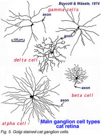
A particularly successful morphological classification scheme was proposed by Boycott and Wassle (1974). Three main classes of physiological response type were correlated with three morphological classes of ganglion cell in cat retina. Thus the alpha, beta, gamma and delta morphological ganglion cell types were considered to be the equivalents of the Y, X and W types of the physiology (Boycott and Wassle, 1974; Enroth-Cugell and Robson, 1966; Cleland and Levick, 1974, Levick and Thibos, 1983, for a review). By comparison with Cajals drawings, it was clear that alpha and beta types were already described (see Cajal drawing of dog and ox retinal ganglion cells) and a further ten or more of Cajals (1892) types were lumped under the umbrella of gamma and delta groups.
By 1978, we knew that alpha and beta cells of Boycott and Wassle (1974) could be subdivided into separate subtypes depending on whether they branch in sublamina a or sublamina b of the inner plexiform layer (Famiglietti and Kolb, 1976) (see chapter on inner plexiform layer). Sublamina a contained dendrites of cells with physiologically defined OFF-center receptive fields and sublamina b dendrites of cell with ON-center receptive fields (Nelson et al., 1978). A new classification of Golgi stained ganglion cells (Kolb et al., 1981) could now relate both alpha and beta cells to the functionally important sublamina scheme and further, described many new types of cell including and going beyond the gamma and delta cells. The 20 cell types over and above the alpha, beta and gamma cells were named from G4-G23 in order of cell body size and dendritic form (i.e. from smallest types, G4 to largest types at G23) (Kolb et al., 1981).
References
- ↑ Kolb, Helga, Nelson, Ralph, Fernandez, Eduardo, Jones, Bryan, The Organization of the Retina and Visual System, Simple Anatomy of the Retina. url=http://webvision.med.utah.edu/book/part-i-foundations/simple-anatomy-of-the-retina/


