Difference between revisions of "The Eye and Retina"
(Created page with "The retina is a light-sensitive layer of tissue that lines the rear surface of the eye. Light from one's visual field passes through the eye and projects onto the retina to cr...") |
Amyrobinson (Talk | contribs) (jin's edits) |
||
| Line 1: | Line 1: | ||
The retina is a light-sensitive layer of tissue that lines the rear surface of the eye. Light from one's visual field passes through the eye and projects onto the retina to create an image. Subsequently, retinal neurons detect this image, which initiates a cascade of biochemical and electrical processing that is sent through the optic nerve and eventually to the visual cortex of the brain. These biochemical and electrical signals provide the basis for vision. | The retina is a light-sensitive layer of tissue that lines the rear surface of the eye. Light from one's visual field passes through the eye and projects onto the retina to create an image. Subsequently, retinal neurons detect this image, which initiates a cascade of biochemical and electrical processing that is sent through the optic nerve and eventually to the visual cortex of the brain. These biochemical and electrical signals provide the basis for vision. | ||
| + | |||
| + | |||
| + | Hey EyeWirers! | ||
| + | |||
| + | As most of us know, we are tracing a sample from the retina of a mouse. Why a mouse and not a human? Why the retina and not a piece of the brain? | ||
| + | |||
| + | We decided to start a blog series with some information on the anatomy and physiology of the human eye and vision. Hopefully you guys will enjoy it. We will start this journey with a simple anatomy of the parts of the eye we. From this point, we will travel inwards the same way light does. Along the way, we will talk about some structures along the way and any other curious information.” | ||
| + | |||
| + | |||
| + | |||
| + | Introduction to the Eye 001/101 - outline | ||
| + | |||
| + | Introduction to what is ahead | ||
| + | |||
| + | |||
| + | Basic anatomy of the human eye | ||
| + | |||
| + | Look into my eyes and tell me what you see | ||
| + | |||
| + | |||
| + | Eyelids and eyelashes | ||
| + | Tear duct | ||
| + | Sclera | ||
| + | White part of the eye | ||
| + | Iris | ||
| + | Color of the eye | ||
| + | Pupil | ||
| + | Dark area at the center of the iris | ||
| + | |||
| + | |||
| + | |||
| + | Image Credits: | ||
| + | http://www.rgbstock.com/bigphoto/mC2Gt1u/eyes+again… | ||
| + | http://m.rgbimg.com/cache1oVsb1/users/g/gr/greyman/600/mC2Gt1u.jpg | ||
| + | http://www.teresewinslow.com/portshow.asp?nxt=13&sid=DD07F1BD-E4CE-4872-984D-A5D8B4A79B60&portfolioid={4B56C61F-9C24-47C6-9F4D-9444E1D75BA2} | ||
| + | |||
| + | More anatomy - Going deeper into the human eye | ||
| + | |||
| + | Situated in the orbit of the skull | ||
| + | |||
| + | |||
| + | Cornea | ||
| + | Transparent surface of the eye that covers the pupil and iris | ||
| + | First refractive surface that light goes through on its way to the retina | ||
| + | 3 layers (epithelium, stroma, endothelium) 5, possibly 6 layers, (Epithelium, Bowmans layer, Stroma, Dua’s layer (Discovered last year, still awaiting official confirmation), Descemet’s membrane, endothelium) | ||
| + | medical: corneal abrasions, ametropia (any refractive area can be cause), etc | ||
| + | Iris | ||
| + | Muscular diaphragm that controls the size of the pupil and amount of light that can enter the eye | ||
| + | Color of the iris is due to quantity and type of melanin | ||
| + | Pupil | ||
| + | Aperture that allows the light to enter the eye | ||
| + | Lens | ||
| + | Second refractive surface that light goes through on its way to the retina | ||
| + | tidbit: no blood supply to the lens | ||
| + | medical: cataracts, presbyopia | ||
| + | Ciliary body | ||
| + | Ciliary muscle (with zonule fibers) | ||
| + | Changes the shape of the lens and allows for accommodation | ||
| + | Ciliary epithelium | ||
| + | Makes the aqueous fluid that fills the anterior and posterior chambers | ||
| + | Three fluid chambers | ||
| + | Anterior | ||
| + | Between cornea and iris | ||
| + | Posterior | ||
| + | Between iris and lens | ||
| + | Vitreous | ||
| + | Between lens and retina | ||
| + | Humours (hehehe) | ||
| + | Aqueous | ||
| + | Clear, watery fluid that supplies nutrients to the structures it surrounds. | ||
| + | Movement of aqueous humor is from posterior chamber to anterior chamber through the pupil. Drainage out of the anterior chamber via canal of Schlemm and trabecular meshwork into venous system. The equilibrium maintains appropriate intra-ocular pressure. | ||
| + | medical: glaucoma (open and close angle) | ||
| + | Vitreous | ||
| + | thick, gel-like fluid that maintains the shape of the eye | ||
| + | 80% of the volume of the eye | ||
| + | medical: floaters, retinal detachment | ||
| + | Three layers of the eye sphere (outside to inside) | ||
| + | Sclera | ||
| + | Outermost layer of the globe | ||
| + | White fibrous layer that becomes transparent at anterior part of the eye and forms the cornea | ||
| + | Choroid | ||
| + | Middle layer between sclera and retina | ||
| + | It is the vascular layer and source of oxygen and nutrients to the outer layers of the retina (photoreceptor layer) | ||
| + | Bruch’s membrane | ||
| + | Retina | ||
| + | Inner layer of the globe | ||
| + | Light-sensitive neurons transmit visual signals | ||
| + | Macula and Fovea | ||
| + | Site of high acuity of vision | ||
| + | Optic nerve (add this here?) | ||
| + | Retinal arteries and veins (add this here?) | ||
| + | |||
| + | Interactive site: http://www.aao.org/theeyeshaveit/anatomy/section-eye.cfm | ||
| + | |||
| + | Extraocular muscles of the eye | ||
| + | |||
| + | |||
| + | Muscles insert into the sclera and move the eyeball and keep image focused on the fovea | ||
| + | 6 main muscles: | ||
| + | superior rectus | ||
| + | inferior rectus | ||
| + | medial rectus | ||
| + | lateral rectus | ||
| + | superior oblique | ||
| + | inferior oblique | ||
| + | medical: lazy eye, etc. | ||
| + | |||
| + | Image Credits: | ||
| + | http://www.ncbi.nlm.nih.gov/books/NBK11120/figure/A726/ | ||
| + | http://www.ncbi.nlm.nih.gov/books/NBK11120/bin/ch11f1.jpg | ||
| + | http://commons.wikimedia.org/wiki/File:1412_Extraocular_Muscles.jpg | ||
| + | http://upload.wikimedia.org/wikipedia/commons/3/3b/1412_Extraocular_Muscles.jpg | ||
| + | Reference Credits: | ||
| + | http://www.ncbi.nlm.nih.gov/books/NBK11120/ | ||
| + | http://www.ncbi.nlm.nih.gov/books/NBK11534/ | ||
| + | |||
| + | |||
| + | |||
| + | |||
| + | |||
| + | Anatomy of the human retina | ||
| + | |||
| + | Funduscopic examination of the retina | ||
| + | |||
| + | |||
| + | Optic cup and disc | ||
| + | retinal ganglion axons converge here | ||
| + | central area are retinal artery and veins | ||
| + | “Blind spot” | ||
| + | Macula and Fovea | ||
| + | high quantity of ganglion cells and cones for visual acuity and color perception | ||
| + | Interactive site: | ||
| + | http://www.aao.org/theeyeshaveit/anatomy/normal-fundus.cfm | ||
| + | Image credits: | ||
| + | http://stanfordmedicine25.stanford.edu/Assets/Images/NormalRetina.gif | ||
| + | |||
| + | |||
| + | 10 histological layers of the retina | ||
| + | http://www.retinalmicroscopy.com/species.html | ||
| + | http://www.retinalmicroscopy.com/movies.html | ||
| + | Retinal pigment epithelium | ||
| + | Single layer of hexagonal cells | ||
| + | Located between the choroid and the photoreceptor layer | ||
| + | Forms a blood-retina barrier with tight junctions with the choroid | ||
| + | It is not firmly attached to the the neural aspect of the retina (photoreceptor layer) | ||
| + | medical: a potential site of retinal detachment | ||
| + | Photoreceptor layer | ||
| + | Composed of rods and cones | ||
| + | Outer limiting “membrane” | ||
| + | Site of connection between photoreceptors and Müller cells | ||
| + | Outer nuclear layer | ||
| + | Nuclei of photoreceptor cells | ||
| + | Outer plexiform layer | ||
| + | Photoreceptor fibers | ||
| + | Bipolar cell dendrites | ||
| + | “Two important synaptic interactions that occur at the outer plexiform layer are: | ||
| + | the splitting of the visual signal into two separate channels of information flow, one for detecting objects lighter than background and one for detecting objects darker that background | ||
| + | the instillation of pathways to create simultaneous contrast of visual objects | ||
| + | In the first synaptic interactions, the channels of information flow are known as the basis of successive contrast, or ON and OFF pathways, respectively, whereas the second interaction puts light and dark boundaries in simultaneous contrast and forms a receptive field structure, with a center contrasted to an inhibitory surround.” | ||
| + | Inner nuclear layer | ||
| + | Bipolar cell nuclei | ||
| + | Horizontal cells | ||
| + | Amacrine cells | ||
| + | Interplexiform cells | ||
| + | Muller cells | ||
| + | Inner plexiform layer | ||
| + | Presynaptic dendrites of bipolar cells (axons) | ||
| + | Postsynaptic dendrites of ganglion cells | ||
| + | Amacrine cell dendrites | ||
| + | Ganglion cell layer | ||
| + | Nerve fiber layer | ||
| + | Axons of Ganglion cells | ||
| + | Inner limiting “membrane” | ||
| + | Ends of Muller cells | ||
| + | |||
| + | |||
| + | |||
| + | |||
| + | Image credit: | ||
| + | http://www.ncbi.nlm.nih.gov/books/NBK11536/figure/ch06ipl.F2/?report=objectonly | ||
| + | http://www.ncbi.nlm.nih.gov/books/NBK11533/figure/ch02sretina.F2/?report=objectonly | ||
| + | http://www.ncbi.nlm.nih.gov/books/NBK10885/figure/A740/?report=objectonly | ||
| + | Reference credit: | ||
| + | http://www.ncbi.nlm.nih.gov/books/NBK10885/ | ||
| + | http://www.ncbi.nlm.nih.gov/books/NBK11533/ | ||
| + | http://www.ncbi.nlm.nih.gov/books/NBK54392/ | ||
| + | http://www.ncbi.nlm.nih.gov/books/NBK11518/ | ||
| + | http://www.ncbi.nlm.nih.gov/books/NBK11536/ | ||
| + | |||
| + | |||
| + | |||
| + | Cells of the retina | ||
| + | Retinal Pigment Epithelium | ||
| + | http://www.retinalmicroscopy.com/pigment.html | ||
| + | Contain pigment granules and absorbs scattered light | ||
| + | Regenerates 11-cis-retinal the chromophore used in photoreceptors | ||
| + | Responds to oxidative stress | ||
| + | Clearing up shed discs of rods and cones | ||
| + | Blood-retinal barrier functions | ||
| + | Medical: | ||
| + | albinos lack pigment in this layer | ||
| + | macular degeneration | ||
| + | retinitis pigmentosa | ||
| + | Photoreceptors | ||
| + | Segments (outer, inner, fiber) | ||
| + | http://www.retinalmicroscopy.com/photoreceptors.html | ||
| + | Rods: role in peripheral vision, night vision | ||
| + | contain rhodopsins | ||
| + | more rods than cones in the retina | ||
| + | no rods in fovea = night blind | ||
| + | more sensitive to dim light | ||
| + | increases in quantity peripherally | ||
| + | Cones: role in visual acuity and color vision | ||
| + | concentrated at center (fovea and macula) and less at periphery | ||
| + | contains different types of opsins | ||
| + | 3 types - each absorb one of 3 colors of light | ||
| + | S-cone: short wavelength - blue | ||
| + | M-cone: medium wavelength - green | ||
| + | L cone: long wavelength -red | ||
| + | |||
| + | |||
| + | Medical: | ||
| + | if issues with one or more cones types - colorblindness | ||
| + | most common - can’t differentiate red and green | ||
| + | usually X-linked recessive - affects men more | ||
| + | http://www.colourblindawareness.org/colour-blindness/ | ||
| + | Ishihara test | ||
| + | Vitamin A deficiency | ||
| + | Retinitis pigmentosa | ||
| + | Dark adaptation (discuss in the future) | ||
| + | Phototransduction (discuss somewhere else in the future) | ||
| + | Bipolar cells | ||
| + | http://www.retinalmicroscopy.com/bipolar.html | ||
| + | Several types | ||
| + | rod-specific bipolar cells (1) | ||
| + | cone-specific bipolar cells (10) | ||
| + | Transmit signals from photoreceptor cells to ganglion cells | ||
| + | processes/neurites are called dendrites | ||
| + | ON and OFF layers | ||
| + | need to explain this* | ||
| + | “We know that a photoreceptor neurotransmitter (which is glutamate, see Dowling (24) and Massey (25) for reviews) is released in the dark in the vertebrate retina (26). Thus, the photoreceptor, whether it be rod or cone, is in a depolarized state in the dark. On light stimulation, the photoreceptor responds with a hyperpolarization; transmitter release ceases, but the postsynaptic bipolar cells respond with either hyperpolarization or depolarization of their membranes. The hyperpolarizing type of bipolar cell is called an OFF-center cell, whereas the depolarizing bipolar cell is called an ON-center cell (27, 28).” | ||
| + | Ganglion cells | ||
| + | http://www.retinalmicroscopy.com/ganglion.html | ||
| + | Bipolar cells contact both dendrites and soma of ganglion cells | ||
| + | Axons → forms optic nerve (CN II) | ||
| + | Association neurons (interneurons) | ||
| + | Modify synaptic transmission in retina | ||
| + | Horizontal cells | ||
| + | http://www.retinalmicroscopy.com/horizontal.html | ||
| + | located between OPL and INL | ||
| + | 3 types on human retina (HI, HII, HIII) | ||
| + | dendrites contact synaptic terminals of photoreceptor cells and with the dendrites of bipolar cells, which they inhibit | ||
| + | |||
| + | Amacrine cells | ||
| + | http://www.retinalmicroscopy.com/amacrine.html | ||
| + | located between INL and IPL | ||
| + | all dendrites emerge from same side of the cell to branch out and terminate in synaptic complexes between bipolar, ganglion, etc cells | ||
| + | thought to lack axons. | ||
| + | some morphologies might have axons but do not leave retina | ||
| + | Interplexiform cells | ||
| + | post-synaptic to amacrine cells and pre-synaptic to horizontal and bipolar cells | ||
| + | feedback loop | ||
| + | Neuroglial cells | ||
| + | cells of Müller - principal glial cells of the retina | ||
| + | http://www.retinalmicroscopy.com/glial.html | ||
| + | extend through the whole thickness of the retina | ||
| + | Provide architectual support | ||
| + | supporting role and other functions (communications?) | ||
| + | Astroglia | ||
| + | Microglia | ||
| + | |||
| + | Image credit: | ||
| + | http://www.as.miami.edu/chemistry/2008-1-MDC/2085/Chap-17_New/chap17_files/image018.jpg | ||
| + | http://media.learn.uci.edu/cat/media/OC08/11004/OC0811004_3RetinalTypes.jpg | ||
| + | http://www.ncbi.nlm.nih.gov/books/NBK11518/figure/A215/?report=objectonly | ||
| + | http://www.ncbi.nlm.nih.gov/books/NBK11536/figure/ch06ipl.F9/?report=objectonly | ||
| + | http://www.as.miami.edu/chemistry/2008-1-MDC/2085/Chap-17_New/chap17_files/image024.jpg | ||
| + | http://www.ncbi.nlm.nih.gov/books/NBK11518/figure/A222/?report=objectonly | ||
| + | http://www.ncbi.nlm.nih.gov/books/NBK11518/figure/A226/?report=objectonly | ||
| + | http://www.ncbi.nlm.nih.gov/books/NBK11536/figure/ch06ipl.F6/?report=objectonly | ||
| + | http://www.ncbi.nlm.nih.gov/books/NBK11516/figure/ch09glia.F2/?report=objectonly | ||
| + | |||
| + | Reference credit: | ||
| + | http://www.ncbi.nlm.nih.gov/books/NBK54392/ | ||
| + | http://www.ncbi.nlm.nih.gov/books/NBK11522/ | ||
| + | http://www.as.miami.edu/chemistry/2008-1-MDC/2085/Chap-17_New/chap17.htm | ||
| + | http://www.ncbi.nlm.nih.gov/books/NBK11518/ | ||
| + | http://www.ncbi.nlm.nih.gov/books/NBK11536/ | ||
| + | http://www.ncbi.nlm.nih.gov/books/NBK11516/ | ||
Revision as of 19:29, 17 April 2014
The retina is a light-sensitive layer of tissue that lines the rear surface of the eye. Light from one's visual field passes through the eye and projects onto the retina to create an image. Subsequently, retinal neurons detect this image, which initiates a cascade of biochemical and electrical processing that is sent through the optic nerve and eventually to the visual cortex of the brain. These biochemical and electrical signals provide the basis for vision.
Hey EyeWirers!
As most of us know, we are tracing a sample from the retina of a mouse. Why a mouse and not a human? Why the retina and not a piece of the brain?
We decided to start a blog series with some information on the anatomy and physiology of the human eye and vision. Hopefully you guys will enjoy it. We will start this journey with a simple anatomy of the parts of the eye we. From this point, we will travel inwards the same way light does. Along the way, we will talk about some structures along the way and any other curious information.”
Introduction to the Eye 001/101 - outline
Introduction to what is ahead
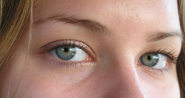
Basic anatomy of the human eye
Look into my eyes and tell me what you see
Eyelids and eyelashes
Tear duct
Sclera
White part of the eye
Iris
Color of the eye
Pupil
Dark area at the center of the iris
Image Credits: http://www.rgbstock.com/bigphoto/mC2Gt1u/eyes+again… http://m.rgbimg.com/cache1oVsb1/users/g/gr/greyman/600/mC2Gt1u.jpg http://www.teresewinslow.com/portshow.asp?nxt=13&sid=DD07F1BD-E4CE-4872-984D-A5D8B4A79B60&portfolioid={4B56C61F-9C24-47C6-9F4D-9444E1D75BA2}
More anatomy - Going deeper into the human eye
Situated in the orbit of the skull
Cornea
Transparent surface of the eye that covers the pupil and iris
First refractive surface that light goes through on its way to the retina
3 layers (epithelium, stroma, endothelium) 5, possibly 6 layers, (Epithelium, Bowmans layer, Stroma, Dua’s layer (Discovered last year, still awaiting official confirmation), Descemet’s membrane, endothelium)
medical: corneal abrasions, ametropia (any refractive area can be cause), etc
Iris
Muscular diaphragm that controls the size of the pupil and amount of light that can enter the eye
Color of the iris is due to quantity and type of melanin
Pupil
Aperture that allows the light to enter the eye
Lens
Second refractive surface that light goes through on its way to the retina
tidbit: no blood supply to the lens
medical: cataracts, presbyopia
Ciliary body
Ciliary muscle (with zonule fibers)
Changes the shape of the lens and allows for accommodation
Ciliary epithelium
Makes the aqueous fluid that fills the anterior and posterior chambers
Three fluid chambers
Anterior
Between cornea and iris
Posterior
Between iris and lens
Vitreous
Between lens and retina
Humours (hehehe)
Aqueous
Clear, watery fluid that supplies nutrients to the structures it surrounds.
Movement of aqueous humor is from posterior chamber to anterior chamber through the pupil. Drainage out of the anterior chamber via canal of Schlemm and trabecular meshwork into venous system. The equilibrium maintains appropriate intra-ocular pressure.
medical: glaucoma (open and close angle)
Vitreous
thick, gel-like fluid that maintains the shape of the eye
80% of the volume of the eye
medical: floaters, retinal detachment
Three layers of the eye sphere (outside to inside)
Sclera
Outermost layer of the globe
White fibrous layer that becomes transparent at anterior part of the eye and forms the cornea
Choroid
Middle layer between sclera and retina
It is the vascular layer and source of oxygen and nutrients to the outer layers of the retina (photoreceptor layer)
Bruch’s membrane
Retina
Inner layer of the globe
Light-sensitive neurons transmit visual signals
Macula and Fovea
Site of high acuity of vision
Optic nerve (add this here?)
Retinal arteries and veins (add this here?)
Interactive site: http://www.aao.org/theeyeshaveit/anatomy/section-eye.cfm
Extraocular muscles of the eye
Muscles insert into the sclera and move the eyeball and keep image focused on the fovea
6 main muscles:
superior rectus
inferior rectus
medial rectus
lateral rectus
superior oblique
inferior oblique
medical: lazy eye, etc.
Image Credits: http://www.ncbi.nlm.nih.gov/books/NBK11120/figure/A726/ http://www.ncbi.nlm.nih.gov/books/NBK11120/bin/ch11f1.jpg http://commons.wikimedia.org/wiki/File:1412_Extraocular_Muscles.jpg http://upload.wikimedia.org/wikipedia/commons/3/3b/1412_Extraocular_Muscles.jpg Reference Credits: http://www.ncbi.nlm.nih.gov/books/NBK11120/ http://www.ncbi.nlm.nih.gov/books/NBK11534/
Anatomy of the human retina
Funduscopic examination of the retina
Optic cup and disc
retinal ganglion axons converge here
central area are retinal artery and veins
“Blind spot”
Macula and Fovea
high quantity of ganglion cells and cones for visual acuity and color perception
Interactive site:
http://www.aao.org/theeyeshaveit/anatomy/normal-fundus.cfm
Image credits:
http://stanfordmedicine25.stanford.edu/Assets/Images/NormalRetina.gif
10 histological layers of the retina
http://www.retinalmicroscopy.com/species.html
http://www.retinalmicroscopy.com/movies.html
Retinal pigment epithelium
Single layer of hexagonal cells
Located between the choroid and the photoreceptor layer
Forms a blood-retina barrier with tight junctions with the choroid
It is not firmly attached to the the neural aspect of the retina (photoreceptor layer)
medical: a potential site of retinal detachment
Photoreceptor layer
Composed of rods and cones
Outer limiting “membrane”
Site of connection between photoreceptors and Müller cells
Outer nuclear layer
Nuclei of photoreceptor cells
Outer plexiform layer
Photoreceptor fibers
Bipolar cell dendrites
“Two important synaptic interactions that occur at the outer plexiform layer are:
the splitting of the visual signal into two separate channels of information flow, one for detecting objects lighter than background and one for detecting objects darker that background
the instillation of pathways to create simultaneous contrast of visual objects
In the first synaptic interactions, the channels of information flow are known as the basis of successive contrast, or ON and OFF pathways, respectively, whereas the second interaction puts light and dark boundaries in simultaneous contrast and forms a receptive field structure, with a center contrasted to an inhibitory surround.”
Inner nuclear layer
Bipolar cell nuclei
Horizontal cells
Amacrine cells
Interplexiform cells
Muller cells
Inner plexiform layer
Presynaptic dendrites of bipolar cells (axons)
Postsynaptic dendrites of ganglion cells
Amacrine cell dendrites
Ganglion cell layer
Nerve fiber layer
Axons of Ganglion cells
Inner limiting “membrane”
Ends of Muller cells
Image credit:
http://www.ncbi.nlm.nih.gov/books/NBK11536/figure/ch06ipl.F2/?report=objectonly
http://www.ncbi.nlm.nih.gov/books/NBK11533/figure/ch02sretina.F2/?report=objectonly
http://www.ncbi.nlm.nih.gov/books/NBK10885/figure/A740/?report=objectonly
Reference credit:
http://www.ncbi.nlm.nih.gov/books/NBK10885/
http://www.ncbi.nlm.nih.gov/books/NBK11533/
http://www.ncbi.nlm.nih.gov/books/NBK54392/
http://www.ncbi.nlm.nih.gov/books/NBK11518/
http://www.ncbi.nlm.nih.gov/books/NBK11536/
Cells of the retina Retinal Pigment Epithelium http://www.retinalmicroscopy.com/pigment.html Contain pigment granules and absorbs scattered light Regenerates 11-cis-retinal the chromophore used in photoreceptors Responds to oxidative stress Clearing up shed discs of rods and cones Blood-retinal barrier functions Medical: albinos lack pigment in this layer macular degeneration retinitis pigmentosa Photoreceptors Segments (outer, inner, fiber) http://www.retinalmicroscopy.com/photoreceptors.html Rods: role in peripheral vision, night vision contain rhodopsins more rods than cones in the retina no rods in fovea = night blind more sensitive to dim light increases in quantity peripherally Cones: role in visual acuity and color vision concentrated at center (fovea and macula) and less at periphery contains different types of opsins 3 types - each absorb one of 3 colors of light S-cone: short wavelength - blue M-cone: medium wavelength - green L cone: long wavelength -red
Medical:
if issues with one or more cones types - colorblindness
most common - can’t differentiate red and green
usually X-linked recessive - affects men more
http://www.colourblindawareness.org/colour-blindness/
Ishihara test
Vitamin A deficiency
Retinitis pigmentosa
Dark adaptation (discuss in the future)
Phototransduction (discuss somewhere else in the future)
Bipolar cells
http://www.retinalmicroscopy.com/bipolar.html
Several types
rod-specific bipolar cells (1)
cone-specific bipolar cells (10)
Transmit signals from photoreceptor cells to ganglion cells
processes/neurites are called dendrites
ON and OFF layers
need to explain this*
“We know that a photoreceptor neurotransmitter (which is glutamate, see Dowling (24) and Massey (25) for reviews) is released in the dark in the vertebrate retina (26). Thus, the photoreceptor, whether it be rod or cone, is in a depolarized state in the dark. On light stimulation, the photoreceptor responds with a hyperpolarization; transmitter release ceases, but the postsynaptic bipolar cells respond with either hyperpolarization or depolarization of their membranes. The hyperpolarizing type of bipolar cell is called an OFF-center cell, whereas the depolarizing bipolar cell is called an ON-center cell (27, 28).”
Ganglion cells
http://www.retinalmicroscopy.com/ganglion.html
Bipolar cells contact both dendrites and soma of ganglion cells
Axons → forms optic nerve (CN II)
Association neurons (interneurons)
Modify synaptic transmission in retina
Horizontal cells
http://www.retinalmicroscopy.com/horizontal.html
located between OPL and INL
3 types on human retina (HI, HII, HIII)
dendrites contact synaptic terminals of photoreceptor cells and with the dendrites of bipolar cells, which they inhibit
Amacrine cells http://www.retinalmicroscopy.com/amacrine.html located between INL and IPL all dendrites emerge from same side of the cell to branch out and terminate in synaptic complexes between bipolar, ganglion, etc cells thought to lack axons. some morphologies might have axons but do not leave retina Interplexiform cells post-synaptic to amacrine cells and pre-synaptic to horizontal and bipolar cells feedback loop Neuroglial cells cells of Müller - principal glial cells of the retina http://www.retinalmicroscopy.com/glial.html extend through the whole thickness of the retina Provide architectual support supporting role and other functions (communications?) Astroglia Microglia
Image credit: http://www.as.miami.edu/chemistry/2008-1-MDC/2085/Chap-17_New/chap17_files/image018.jpg http://media.learn.uci.edu/cat/media/OC08/11004/OC0811004_3RetinalTypes.jpg http://www.ncbi.nlm.nih.gov/books/NBK11518/figure/A215/?report=objectonly http://www.ncbi.nlm.nih.gov/books/NBK11536/figure/ch06ipl.F9/?report=objectonly http://www.as.miami.edu/chemistry/2008-1-MDC/2085/Chap-17_New/chap17_files/image024.jpg http://www.ncbi.nlm.nih.gov/books/NBK11518/figure/A222/?report=objectonly http://www.ncbi.nlm.nih.gov/books/NBK11518/figure/A226/?report=objectonly http://www.ncbi.nlm.nih.gov/books/NBK11536/figure/ch06ipl.F6/?report=objectonly http://www.ncbi.nlm.nih.gov/books/NBK11516/figure/ch09glia.F2/?report=objectonly
Reference credit: http://www.ncbi.nlm.nih.gov/books/NBK54392/ http://www.ncbi.nlm.nih.gov/books/NBK11522/ http://www.as.miami.edu/chemistry/2008-1-MDC/2085/Chap-17_New/chap17.htm http://www.ncbi.nlm.nih.gov/books/NBK11518/ http://www.ncbi.nlm.nih.gov/books/NBK11536/ http://www.ncbi.nlm.nih.gov/books/NBK11516/