Translations:J-RGC/31/en
The pattern by which retinal ganglion cells develop in the retina varies by subtype. It has been found that J-RGCs develop via a gradual restriction of a diffuse pattern. The dendrites of J-RGC cells have been found to extend throughout the entire inner plexiform layer at P5 in mice. At P8 in mice it was found that branches had been pruned in the inner portion of the inner plexiform layer, while in the outer portion of the Inner Plexiform Layer the branches had propagated. In fact, they were found to have their arbors centered around starburst amacrines in SL3. Gradually the arbor distributions were found to shift outward through the IPL until P12, when they had achieved their adult pattern of restriction to LS2.[1]
- Error creating thumbnail: Unable to save thumbnail to destination
The development of retinal ganglion cells varies by cell type. Here we see J-, BD-, W3, and W7-RGCs at stages P5, P8, and P12/13. Note the J-RGC's gradual shifting of dendritic arbor distribution to the outer portion of the IPL.[1]
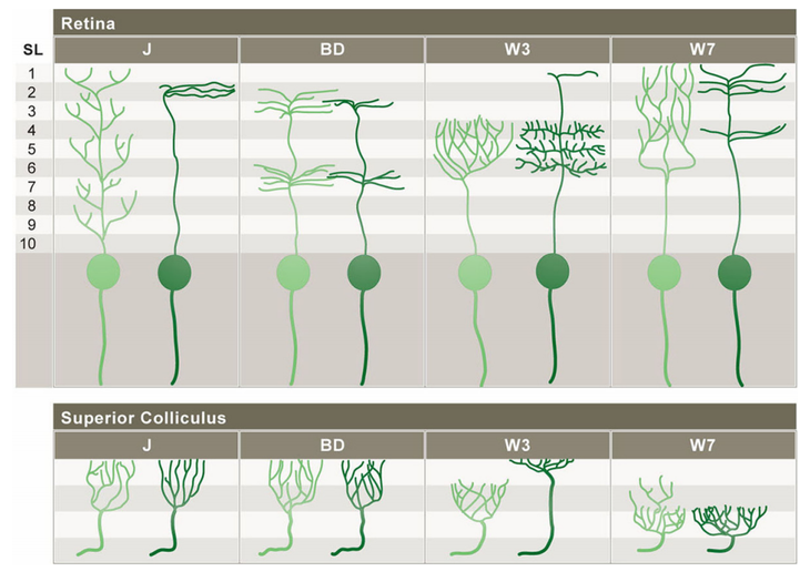
A visual overview of subtype-specific patterns of development in J-, BD-, W3-, and W7-RGCs. The light green figures represent RGC patterns at an immature age and the dark green figures represent the mature patterns. In addition to having specific patterns of dendritic lamination, retinal ganglion cell subtypes have distinctive axonal arbor patterns within the superior colliculus and lateral genticulate nucleus.[1]

- ↑ Cite error: Invalid
<ref>tag; no text was provided for refs namedkim2010